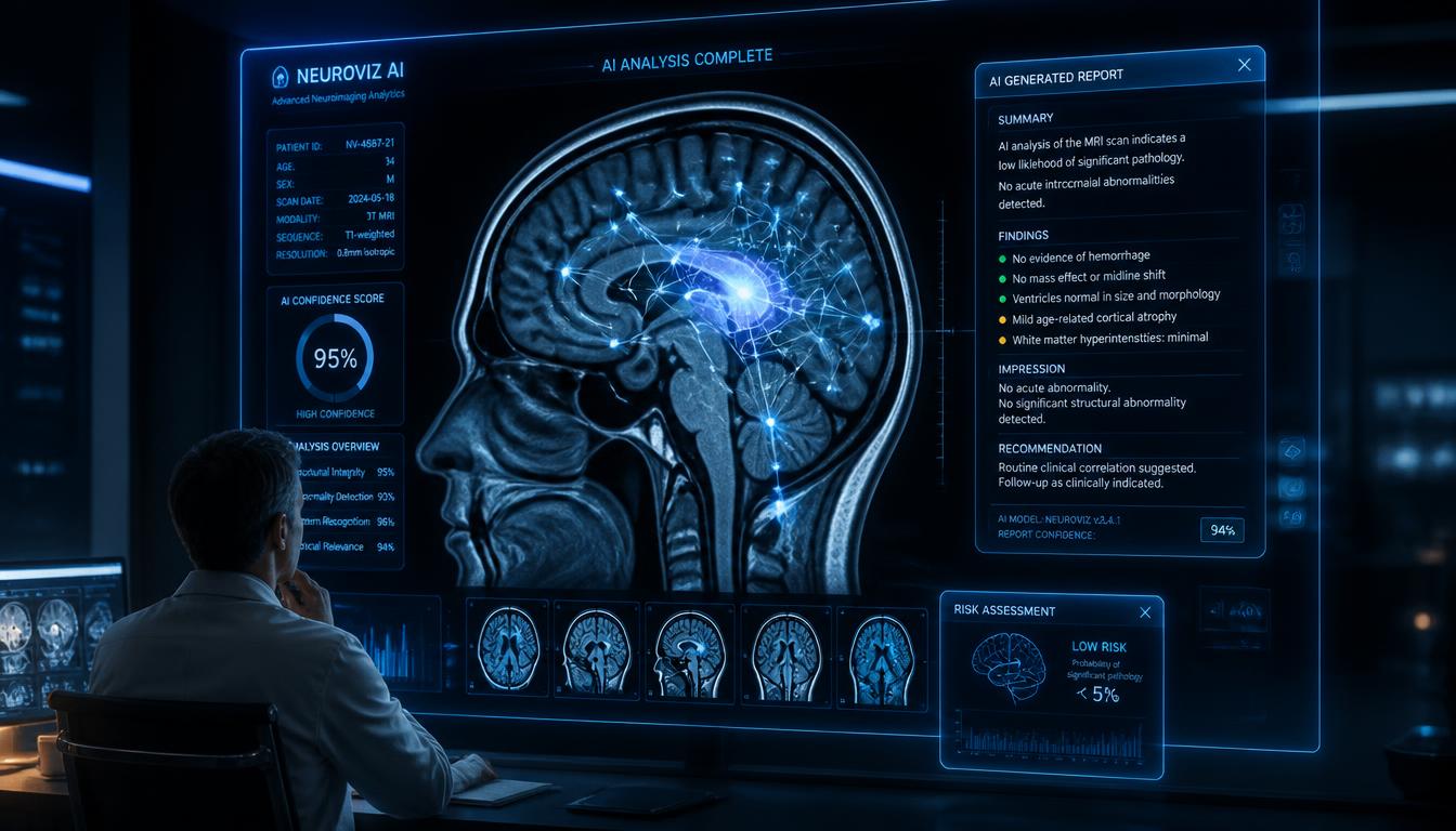
Radiology departments face a productivity crisis: global radiologist workloads have grown 30% in the past decade while radiologist supply has remained flat. The average radiologist reads 50–100 studies daily under growing time pressure. AI radiology report generation addresses this challenge by providing intelligent report drafting assistance, critical finding detection, and structured template completion that allows radiologists to focus on diagnostic judgment rather than documentation mechanics.
How AI Radiology Reporting Works
AI radiology systems analyze medical images (X-rays, CT scans, MRIs, ultrasounds) and generate preliminary structured report drafts that radiologists review and finalize. The AI identifies findings, suggests impression statements, and flags potential critical findings requiring urgent communication. Radiologists spend 30–50% less time on routine report generation while maintaining full clinical oversight of every signed report.
Structured Report Templates
AI-assisted reporting uses structured templates that ensure complete, consistent documentation across modalities, chest CT, brain MRI, abdominal ultrasound, musculoskeletal X-rays, following radiology society standards (ACR, RSNA reporting templates). Structured reports with standardized terminology improve communication to referring physicians, reduce misinterpretation, and enable downstream data analytics that are impossible with free-text reports.
Critical Finding Detection and Notification
AI identifies potentially critical findings (pneumothorax, pulmonary embolism, intracranial hemorrhage, aortic dissection, malignant-appearing masses) and immediately flags them for radiologist priority review. Critical finding notification workflows ensure urgent results reach referring providers within established time standards, automatically documenting the notification for compliance purposes.
Incidental Finding Management
Radiology reports frequently identify incidental findings that require follow-up (adrenal nodules, pulmonary nodules, aortic dilation). AI systems track these findings against established follow-up guidelines (Fleischner Society nodule guidelines, ACR Incidental Findings Committee recommendations), generating automatic follow-up recommendations and tracking whether ordered follow-up studies are completed.
Performance by Modality
Chest X-ray: AI achieves performance comparable to general radiologists for detecting pneumonia, pneumothorax, cardiomegaly, and pleural effusion. Chest CT: Lung nodule detection and characterization. Mammography: Calcification detection and mass characterization assistance. Brain MRI: White matter lesion quantification and volumetric analysis. Each modality requires separate model training and validation.
Turnaround Time Impact
Radiology departments using AI report assistance reduce turnaround times by 30–50% for routine studies. Emergency radiology benefits most, AI pre-reads trauma CTs while the patient is still in the scanner, ensuring findings are ready before the radiologist sits to formally read. Overnight and weekend reads with AI assistance maintain quality without requiring round-the-clock radiologist staffing at smaller facilities.
Integration with RIS and PACS
AI reporting integrates with the Radiology Information System (RIS) for worklist management and with PACS for image access. Reports auto-populate into the RIS from AI analysis, requiring only radiologist review and electronic signature. HL7 FHIR messaging delivers finalized reports to ordering physicians' EMRs within minutes of signature.
Regulatory Considerations
In the USA, AI radiology tools that influence clinical decision-making require FDA 510(k) clearance or De Novo authorization as Software as a Medical Device (SaMD). In the EU, CE marking under the EU Medical Device Regulation (MDR) is required. Hospitals should verify regulatory status before deploying AI radiology tools in clinical production environments.
Ready to optimize your Ai Radiology Report Generation Imaging D workflows? Book a tailored Quecorex demo today.
Human Sign-Off Is Essential
AI can draft impressions, pre-populate templates, and highlight findings for review, but the radiologist remains responsible for the final report. Configure workflows so no AI-generated text is released without radiologist review and signature, and record which parts were drafted by AI. Measure how often radiologists edit the drafts and which findings are most often changed.
Evaluating an AI Imaging Tool
- Check the intended use. Which modality, body region, and findings is it designed for?
- Ask for evidence. Published or independent validation, and performance on data similar to yours.
- Confirm regulatory status. Many AI imaging tools are regulated as medical devices, and status differs by country.
- Test on your own studies. Compare sensitivity and false alarms on a sample of your historical cases.
- Pilot with monitoring. Run in shadow or advisory mode first and track outcomes.
- Plan for drift. Performance can change when equipment, protocols, or populations change.
Questions About Data
- Where are images processed, and do any leave our environment?
- Are images or reports used to train the vendor's models, and under what agreement?
- How is access logged and audited?
- What happens to data if we end the contract?
The underlying workflow is covered in our RIS guide. AI features have no monthly module fee; you buy AI credits and pay for what you use. See the pricing estimator for the modules that do carry a fee.
Where Radiology AI Fits in the Workflow
| Stage | Possible AI role |
|---|---|
| Before the scan | Protocol suggestions, scheduling support, dose optimisation |
| During acquisition | Image quality checks and prompts to repeat poor images |
| Triage | Prioritising studies with suspected urgent findings on the reading list |
| Interpretation | Highlighting regions of interest or measurements for the radiologist |
| Reporting | Drafting text from templates and findings, for review |
| After reporting | Follow-up tracking for incidental findings |
Each role has different risks and evidence. A triage tool that reorders a worklist affects different things than a tool that drafts an impression.
Change Management for Radiologists
- Involve radiologists in selection and configuration from the start.
- Be clear about who is responsible for what the tool shows.
- Provide training on strengths, limits, and known failure modes.
- Collect feedback on missed cases and false alerts and share it with the vendor.
- Make it easy to switch a feature off if it is not helping.
Choosing Your First Use Case
A practical first project has a clear problem, measurable results, and modest risk. Examples include prioritising suspected critical findings on chest images in a busy emergency department, or structuring reports for common examinations. Measure turnaround times, agreement with radiologist findings, and radiologist satisfaction before and after. Connect the tool to the core imaging systems described in our RIS guide and business side in the diagnostic centre guide. Imaging devices should be maintained and calibrated, as covered in biomedical equipment management.
Final Thoughts
AI radiology reporting is one of the most mature and validated areas of clinical AI, with multiple FDA-cleared algorithms commercially available. The technology does not replace radiologists, it amplifies their capabilities, enabling radiologists to read more studies, catch more findings, and communicate results faster than manual workflows allow. Quecorex AI Radiology integrates with any PACS and RIS for seamless deployment in imaging departments worldwide.